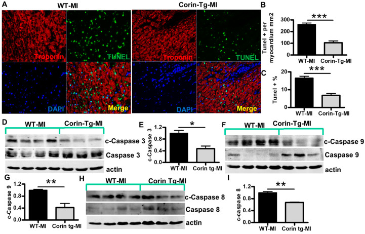
Figure 3.
Cardiac-specific overexpression of corin attenuates cardiomyocyte apoptosis 24 h post-MI. (A) Representative images of TUNEL staining (green) with troponin (red, cardiac marker) and DAPI-stained nuclei (blue) in the ischemic area of left ventricles (LV) from WT and corin-Tg mouse hearts post-MI (40× magnification). (B,C) Quantitative digital analysis of TUNEL staining. The total numbers TUNEL-stained cells (TUNEL+) and DAPI-stained nuclei (total cell) in the LV were counted; the total left ventricular area (LVA) was measured at 5× using Image-Pro Plus software. The ratios of TUNEL+ cells per myocardial area (mm2) and the percentages of TUNEL+ cells vs. total cells were calculated. Data represent means ± SE of n = 4 mice per group. (D,F,H) Western blot analysis of tissue lysates, prepared using the infarct core and border zone myocardium from corin-Tg-MI (n = 3) and WT-MI (n = 4) hearts, with antibodies for cleaved (c) caspase 3 (17 kD), caspase 3 (34 kD) c-caspase 9 (37, 39 kD), caspase 9 (37 kD), c-caspase 8 (18 kD), and caspase 8 (55 kD) under reducing conditions. Actin (43 kD) was used as loading control. (E,G,I) Bar graphs represent the densitometry analysis of c-caspase 3, 9 and 8 normalized to actin. Data represent means ± SE of n = 3–4 mice per group. * p < 0.05, ** p < 0.01, *** p < 0.001.

