. 2020 Jun 8;43(3):e20190153. doi: 10.1590/1678-4685-GMB-2019-0153

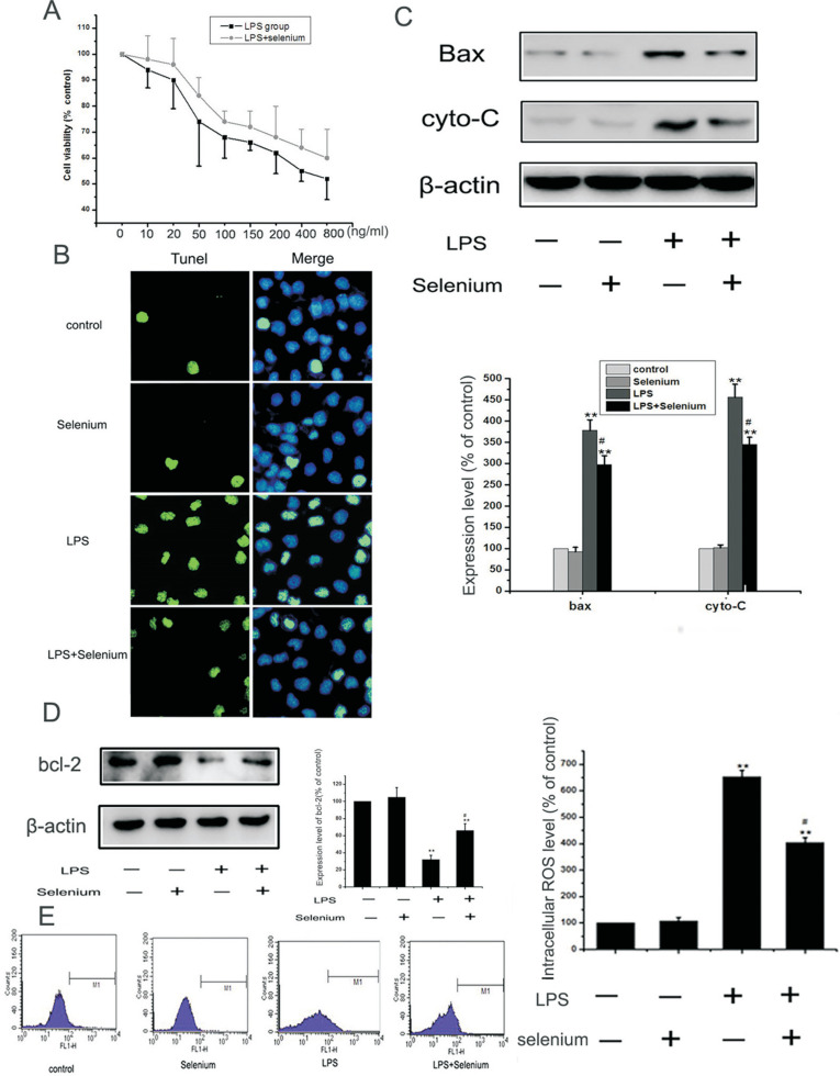
Figure 2. Selenium inhibited LPS-induced apoptosis and oxidative stress in MC3T3-E1 cells. (A) We incubated MC3T3-E1 cells with 0-800 ng/ml of LPS with or without 4 ng/ml sodium selenite for 48 h, and cell viability was detected by MTT. The data were mean ± SEM (n=5). (B) MC3T3-E1 cells were incubated in 200 ng/ml LPS in the presence or absence of 4 ng/ml sodium selenite for 24 h, and the apoptosis was assessed by using TUNEL analysis. (C,D) Cells were treated as above, western blots were performed with the antibodies indicated. Relative expression of Bax, cytochrome c and bcl-2 were calculated and normalized to the loading control β-actin. The data were mean ± SEM (n = 6). (** P < 0.01, vs. control, #P< 0.05, LPS vs. LPS+selenium group). (E): Cells were treated as aboved, and the level of intracellular ROS production were detected by FACS analysis. The data were mean ± SEM (n = 3). (** P < 0.01, vs. control, #P< 0.05, LPS vs. LPS+selenium group).

Figure 2