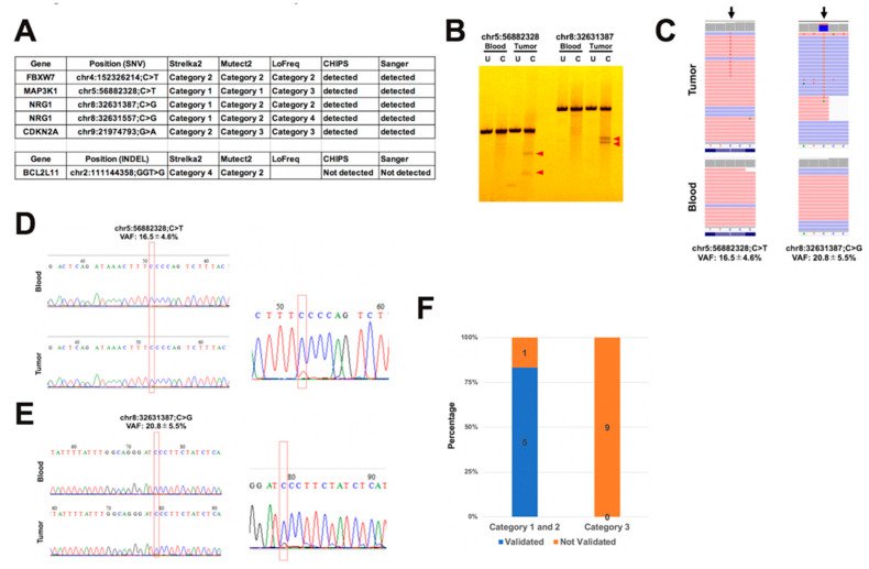
Figure 4.
Validation for the detected somatic variants by CHIPS and Sanger sequencing. (A) Summary of validation results. (B) CHIPS technology assay. Red arrow head shows cleaved heteroduplexes. U: uncut C: cut by CEL nuclease. (C) Aligned reads visualized by Integrative Genomic Viewer (IGV) at the detected somatic variant positions. (D) and (E) Electropherograms of Sanger sequencing of the detected somatic variants in blood and tumor samples. (F) Ratio of validated and not validated variants in category 1, 2, and 3.

