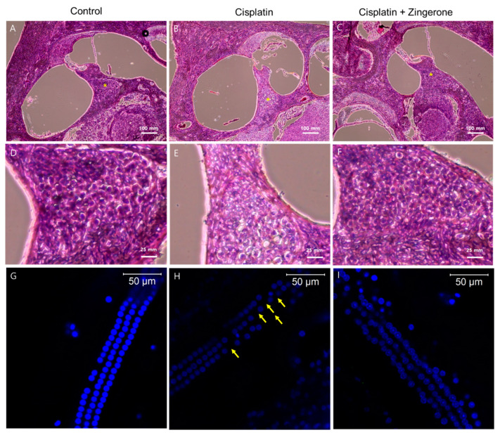
Figure 4.
Hematoxylin and eosin (H&E) staining of the cochlea (A–F) and whole mounts of the cochlear basal turn (G–I). The control group exhibited intact spiral ganglion cells (A) and outer hair cells (G). Compared with the cisplatin group (B,E,H), the cisplatin + zingerone group (C,F,I) exhibited a preserved number and arrangement of spiral ganglion cells (* in A–C and D–F) and outer hair cells (the arrows indicate the loss of outer hair cells).

