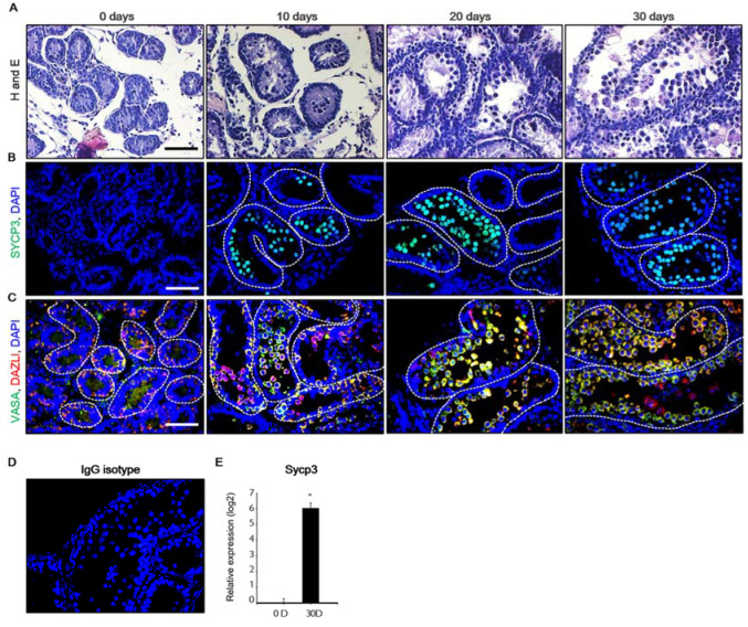
Figure 1.
Development of mouse testicular fragments (MTFs) in the in vitro culture model. (A) Histological assessments performed using hematoxylin and eosin staining of MTFs cultured for 0, 10, 20, and 30 days. (B) SYCP3, (C) VASA, and DAZL proteins were detected in the MTFs after 0, 10, 20, and 30 days of culture using immunostaining. (D) The negative control stain using isotype-matched IgGs showed no specific signal. (E) The mRNA levels of the meiotic marker Sycp3 in the MTFs were examined using quantitative polymerase chain reaction analysis. The relative quantification of mRNA is shown using the mean and standard error of the mean (n = 6) at log2 scale. * p < 0.05, Scale bars = 50 μm; each image was observed at the same magnification.

