Figure 3.
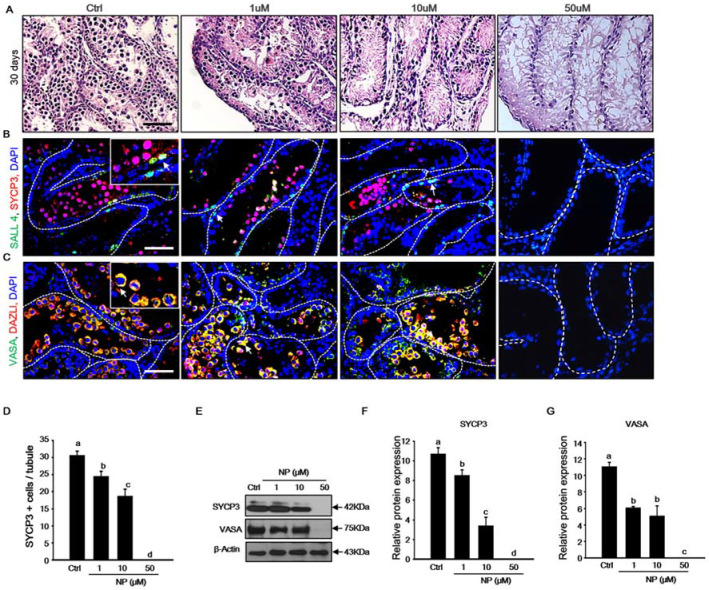
Toxic effect of nonylphenol (NP) on germ cell development. (A) Histological features of the mouse testicular fragments (MTFs) cultured for 30 days with 0, 1, 10, and 50 μM NP. (B) Meiotic and undifferentiated germ cells co-stained with SYCP3 and SALL4 antibody to confirm the occurrence of meiosis and the survival of undifferentiated germ cells in NP-exposed MTFs. SYCP3- and SALL4-positive cells (white arrow) were observed in 0, 1, and 10 μM NP-treated MTFs, but not in the 50 μM NP-treated MTFs. (C) MTFs co-stained with the germ cell markers VASA and DAZL in the presence and absence of NP (0, 1, 10, and 50 μM). The white arrow indicates VASA- and DAZL-positive cells in the germinal epithelium, and these cells were evident in 0, 1, and 10 μM NP-treated MTFs, but not in the 50 μM NP-treated MTFs. Scale bars = 50 µm. All images were acquired at the same magnification. (D) The average number of differentiated germ cells per seminiferous tubule was calculated on the basis of SYCP3 immunostaining in the 0, 1, and 10 μM NP-treated MTFs. At least 50 tubules were scored for each MTF (5–6 biological replicates). The data are shown as mean ± standard error. (E) The levels of SYCP3 and VASA proteins were measured in the MTF lysate with or without NP treatment, and β-actin was used as a loading control. The relative expression of (F) SYCP3 and (G) VASA in the MTF lysates is shown using the mean and the standard error of the mean (n = 5).

