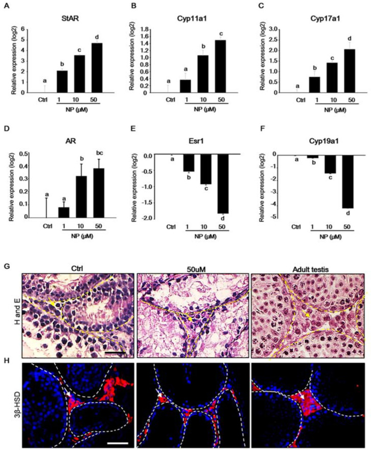
Figure 5.
Expression of Leydig cell markers in the mouse testicular fragments (MTFs) after nonylphenol (NP) treatment. MTFs were cultured in the absence and presence of NP (1–50 μM) for 30 days. (A) Star, (B) Cyp11α1, (C) Cyp17a1, (D) Ar, (E) Esr1, and (F) Cyp19α1 mRNA levels in the MTFs were determined using quantitative polymerase chain reaction. The relative quantification of mRNA is shown as the mean and the standard error of the mean (n = 6) at log2 scale. The expression of steroidogenesis-related Leydig cell markers such as Star, Cyp11α1, and Cyp17a1 increased dose-dependently in the NP-treated MTFs. (G) The histological image shows that Leydig cells are located in the interstitial region in NP-treated (50 μM) and untreated MTFs (indicated by yellow arrow), and adult testes were used as positive controls. Moreover, (H) 3β-HSD protein was detected in MTFs with and without NP treatment by immunostaining (indicated by white arrow). Scale bars = 50 µm. All of the images were acquired at the same magnification.

