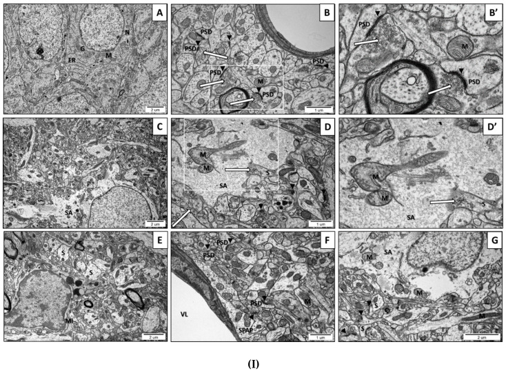
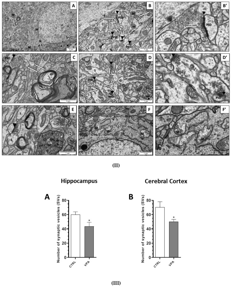
Figure 2.
(I). The effect of prenatal exposure to VPA on the ultrastructure of neuronal cells in the CA1 region of the hippocampus of the offspring. (A–B’) Control group. Ultrastructurally unchanged neuronal cells, unaltered structure of neuropil with normal appearance of the synaptic cleft (black arrowheads), well-defined structure of the synapses with accurate postsynaptic density (PSD), the correct distribution of synaptic vesicles (SVs) (long arrows), and ultrastructurally unchanged mitochondria (M). (C–G) VPA-exposed group. Reduced packing density of SVs in the presynaptic area (release of SVs from the presynaptic area accompanied by disruption of the synaptic membranes) (long arrow), nerve ending swelling (S), blurred and thickened structure of the synaptic cleft without clearly marked pre- and postsynaptic membranes (black arrowheads). Ultrastructurally changed mitochondria with a blurred cristae structure (M). Astrocytes with features of swelling (SA) and swollen perivascular astrocyte processes (SPAP) were observed. Microglial cell activation (Mi); (VL) Vascular lumen; (G) Golgi apparatus; (ER) Endoplasmatic reticulum; (N) Neuron. Representative pictures from n = 6 independent experiments for the control and experimental animals are presented. (II). The effect of prenatal exposure to VPA on the ultrastructure of neuronal cells in the cerebral cortex of the offspring. (A–B’) Control group. Ultrastructurally unchanged neuronal cells, unaltered structure of neuropil with normal appearance of the synaptic cleft (black arrowheads), well-defined structure of synapses with accurate postsynaptic density (PSD), correct distribution of SVs (long arrows), and well preserved mitochondria (M). (C–F’) VPA-exposed group. Reduced packing density of SVs in the presynaptic area (release of SVs from the presynaptic area accompanied by the disruption of the synaptic membranes) (long arrows), nerve ending swelling (S), and blurred and thickened structure of the synaptic cleft, without clearly marked pre- and postsynaptic membranes (black arrowheads). Ultrastructurally changed mitochondria with a blurred cristae structure (M). Changed myelin structure (CHM); (DN) Degenerating neuron; (N) Neuron. Representative pictures from n = 6 independent experiments for the control and experimental animals are presented. (III). The effect of prenatal exposure to VPA on the synaptic vesicles (SVs) number. The effect of VPA on the number of synaptic vesicles (SVs) in the CA1 region of the hippocampus (A) and the cerebral cortex (B) was analysed at postnatal day 58 (PND58). The number of SVs was counted in 30 nerve endings of each animal from the control and experimental groups. Data represent the mean values ± SEM from n = 4 independent experiments. * p ˂ 0.05, vs. control.


