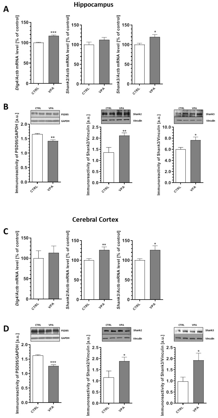
Figure 5.
The effect of prenatal exposure to VPA on the expression of the postsynaptic density proteins: PSD95, Shank2, and Shank3. The gene expression of Dlg4, Shank2, and Shank3 in the hippocampus (A) and cerebral cortex (C) of the control and VPA-exposed rats was measured by quantitative RT-PCR and calculated by the ΔΔCt method with Actb (β-actin) as a reference gene. Data represent the mean values ± SEM from n = (4–5) independent experiments in the hippocampus and n = (4–8) in cortex. The immunoreactivity of the postsynaptic density proteins in the control and VPA-exposed rats was monitored using Western blot analysis. Densitometric analysis and representative pictures for PSD95, Shank2, and Shank3 in the hippocampus (B) and cerebral cortex (D) are presented. The results were normalized to GAPDH or vinculin levels. Data represent the mean values ± SEM from n = (4–9) independent experiments in both the hippocampus and cerebral cortex. * p < 0.05, ** p < 0.01, *** p < 0.001 vs. control.

