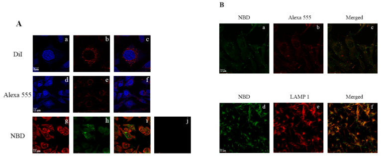
Figure 7.
Lysosomal targeting of rHDL/res/NBD by apoE3 in glioblastoma cells. (A) Uptake of individual components of rHDL/res was monitored by direct or indirect fluorescence: lipid (a–c), apoE3 (d–f) and resveratrol (g–i). Following exposure to rHDL/res/DiI at 37 °C for 3 h (a–c), the cells were visualized under a confocal laser scanning microscope: (a) DAPI; (b) DiI; (c) merge of (a) and (b). Following exposure to rHDL/res under the same conditions (d–f), the cells were visualized by: (d) DAPI; (e) apoE3 monoclonal antibody, 1D7, and Alexa555-conjugated secondary antibody; (f) merge of (d) and (e). Following exposure to rHDL/res/NBD (5 μg) as described above (g–i), the cells were visualized by: (g) propidium iodide; (h) resveratrol conjugated to 4-chloro-7-nitrobenz-2-oxa-1,3-diazole (res/NBD); (i) merge of (g) and (h). Panel (j) shows that uptake of res/NBD is minimal in the absence of rHDL. (B) Co-localization of res/NBD with apoE3, or with LAMP1 in late endosomal/lysosomal vesicles following cellular uptake of rHDL/res/NBD. Following exposure to rHDL/res/NBD, the cells were visualized by fluorescence associated with: (a) NBD to detect res, (b) Alexa555-conjugated secondary antibody to detect apoE3; (c) merge of (a) and (b); (d) NBD to detect res; (e) Alexa 594-conjugated secondary antibody to detect LAMP1; (f) merge of (d) and (e). Reproduced with permission from [63]. Copyright Public Library of Science, 2015.

