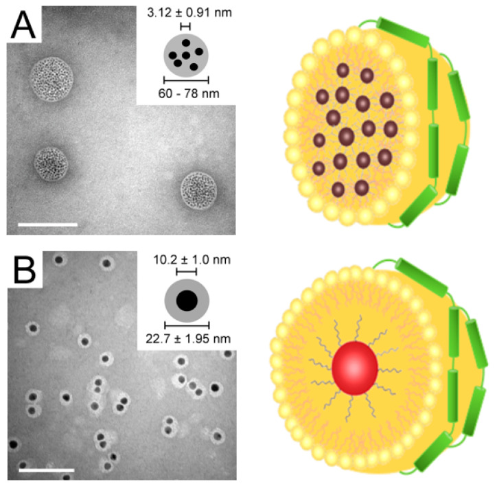
Figure 8.
Nanolipoproteins reconstituted with 3 nm and 10 nm AuNP. TEM image (Left) and schematic representation (Right) of 3 nm (A) and 10 nm (B) AuNP incorporated into rHDL/apoE3. The scale bar represents 100 nm in TEM images. TEM image of rHDL-AuNP prepared with 3 nm AuNP (A) revealed 60–80 nm spheroid structures with several AuNP (Inset, A), while that prepared with 10 nm AuNP revealed ~23 nm spherical structures with a single AuNP (Inset, B). The light area around the 10 nm AuNP likely represents the lipoprotein shell. Reproduced with permission from [64]. Copyright Dove Medical Press, 2017.

