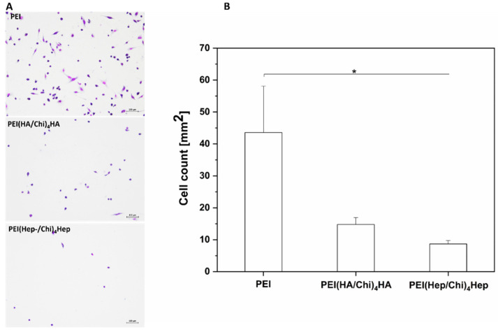
Figure 4.
(A) Transmitted light microscopy images of adherent macrophages stained with 10% (v/v) Giemsa after 24 h on poly (ethylene imine) (PEI) and terminal layers of PEMs composed of either hyaluronic acid (HA) or heparin (Hep) as polyanions and chitosan (Chi) as polycation abbreviated as (PEI(HA/Chi)4HA, PEI(Hep/Chi)4Hep), respectively. Scale: 100 μm. (B) Number of adherent macrophages per surface area after 24 h of cultivation. Data represent means ± SD, n = 5, * p ≤ 0.05.

