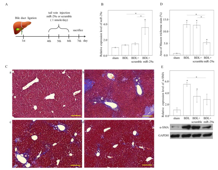
Figure 1.
Exogenous miR-29a injection significantly reduces liver fibrosis in the context of BDL. (A) Experimental procedure. (B) quantitative real-time PCR (qRT-PCR) results of miR-29a levels in liver specimens. N = 6–13. (C) Representative image of Masson trichrome staining. a: sham, b: BDL, c: BDL + scramble, d: BDL+miR-29a. Blue stain indicates collagen matrix accumulation. Scale bar, 200 μm(D) quantification results of Masson trichrome staining. Positive staining area (%) was quantified using ImageJ. N = 6–7. (E) Representative blotting image and densitometric results of α-SMA protein expression. N = 6 for each group. Histogram data are expressed as mean ± SE. * p < 0.05 between the groups. Sham, sham surgery only. BDL, bile duct ligation operation only. BDL + scramble, mice received exogenous scramble injection after BDL. BDL + miR-29a, mice received exogenous miR-29a injection after BDL. α-SMA, alpha-smooth muscle actin.

