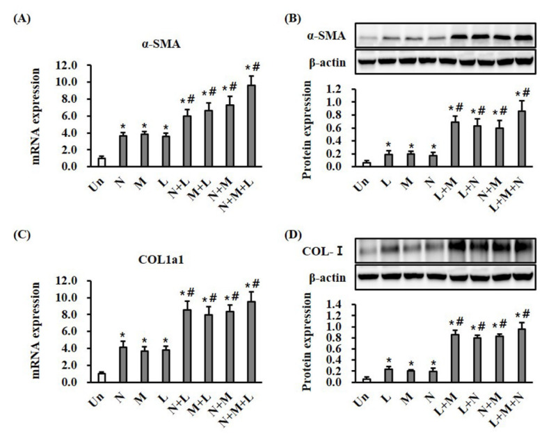
Figure 4.
Mono- and combination therapy increased α-SMA expression and collagen deposition in wound tissues. (A) Results of RT-qPCR analysis of α-SMA in wound tissue after treatment for 7 days. The mRNA expression levels are displayed as a fold change with respect to the levels in the control by the 2−ΔΔCt method. * p < 0.05 vs. the control group, and # p < 0.05 vs. the control and comparable monotherapy groups. (B) Results of Western blotting analysis of α-SMA in wound tissue. The protein expression levels were normalized with β-actin. * p < 0.05 vs. the control group, and # p < 0.05 vs. the control and comparable monotherapy groups. (C) Results of RT-qPCR analysis of type I collagen in wound tissue. The mRNA expression levels are displayed as a fold change with respect to the levels in the control by the 2−ΔΔCt method. * p < 0.05 vs. the control group, and # p < 0.05 vs. the control and comparable monotherapy groups. (D) Results of the Western blotting analysis of type I collagen in wound tissue. The protein expression levels were normalized with β-actin. * p < 0.05 vs. the control group, and # p < 0.05 vs. the control and comparable monotherapy groups. Data are mean ± SEM (n = 5). LTP, low-temperature plasma; NPWT, negative pressure wound therapy; MSC, bone marrow mesenchymal stem cell; and Un, control.

