Figure 4.
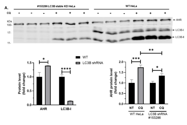
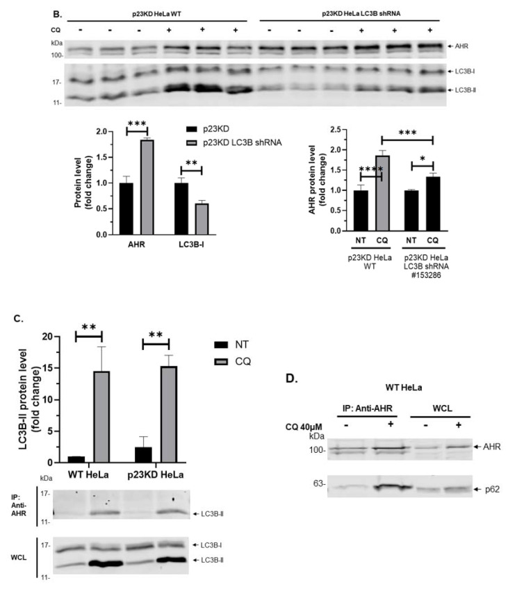
AHR interacts with LC3B-II and p62 in p23 stable knockdown (p23KD) and wild type (WT) HeLa cells. (A) Stable knockdown of LC3B in HeLa cells increased AHR protein levels and reduced the extent of the CQ-mediated increase of AHR. The images above show LC3B-I, LC3B-II, and AHR protein levels in WT and LC3B stable knockdown (KD) HeLa cells with or without treatment of 40 μM CQ for 6 h of triplicate samples. The graphs represent replicate data of means ± SD of one experiment, n = 3. WT (left graph) and NT (right graph) were arbitrarily set as 1 for data normalization. This experiment was repeated once with similar results. Data of the left graph were analyzed by multiple t-tests corrected with the Holm-Sidak method for multiple comparisons to determine statistical significance whereas data of the right graph were analyzed by one-way ANOVA with Sidak’s multiple comparisons test to determine statistical significance. (B) Transient knockdown of LC3B in p23 stable knockdown (p23KD) HeLa cells showed a higher increase of AHR protein levels when compared to that in WT HeLa cells (see A) and reduced the extent of CQ-mediated increase of AHR. The graphs represent replicate data of means ± SD of one experiment, n = 3. p23KD (left graph), and no addition as no treatment (NT, right graph) were arbitrarily set as 1 for data normalization. This experiment was repeated once with similar results. Data of the left graph were analyzed by multiple t-tests corrected with the Holm-Sidak method for multiple comparisons to determine statistical significance whereas data of the right graph were analyzed by one-way ANOVA with Sidak’s multiple comparisons test to determine statistical significance. (C) LC3B-II was co-immunoprecipitated by AHR polyclonal antibody SA210 in both cell lines after treatment of 40 μM CQ for 6 h. Data were presented from three independent experiments as means ± SD, n = 3. WT HeLa NT group was arbitrarily set as one for comparison (no error bar). Data were analyzed by multiple t-tests corrected with the Holm-Sidak method for multiple comparisons to determine statistical significance. The images below are representative of the replicate data. (D) p62 was co-immunoprecipitated by AHR monoclonal antibody A-3x after treatment of 40 μM CQ for 6 h in WT HeLa cells. This experiment was repeated once with similar results. For A-B, each Western lane contained 30 μg of whole-cell lysate or the whole immunoprecipitation content from 1 to 2 mg of whole-cell lysate starting material (C and D). Data were normalized by total protein stain.


