Figure 1.
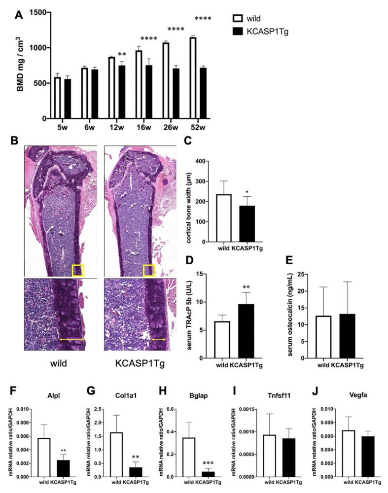
Bone mineral density (BMD) and expression of mRNAs associated to bone formation or destruction in the femur of KCASP1Tg and wild-type mice. BMD was scanned at the distal end of the right femur in KCASP1Tg and wild-type littermates of 5 to 52 weeks of age (n = 4 per group). In KCASP1Tg mice, BMD decreased significantly compared to wild-type littermates at 12 weeks of age (A). The cortical bone width was measured in HE (upper panel × 40, lower panel × 100). Four parts (n = 3 per group) were randomly selected and measured by using Analysis Application Hybrid cell count. The cortical width of the femur was significantly thinner in KCASP1Tg mouse, as determined by histological HE staining (B,C). Bone remodeling markers including TRACP-5b and osteocalcin were measured in the serum by a specific ELISA kit. The serum level of TRACP-5b is a sensitive marker of bone resorption, and was significantly increased in KCASP1Tg mice (D), but the biomarker of bone formation, serum osteocalcin, was unchanged ((E), n = 8 per group). The expression of relevant mRNAs in the right femur was quantified by real time PCR, and the values were standardized by using GAPDH (n = 6 per group). The expressions of genes involved in bone formation, such as alkaline phosphatase-(Alpl, (F)), collagen type I (Col1a1, (G)) and bone gamma-carboxyglutamate protein (Bglap, osteocalcin, (H)) were significantly decreased in KCASP1Tg mice. The mRNA levels of genes for osteoclast differentiation and activation, i.e., members of the tumor necrosis factor superfamily (Tnfsf11, RANKL, (I)) and vascular endothelial growth factor (Vegfa, (J)) were unchanged in the model mice. All data are expressed as mean ± SD. *; p < 0.05, **; p < 0.01, ***; p < 0.001, ****; p < 0.0001 between KCASP1Tg and wild-type mice by Mann–Whitney (except A) test and ordinary one-way ANOVA.

