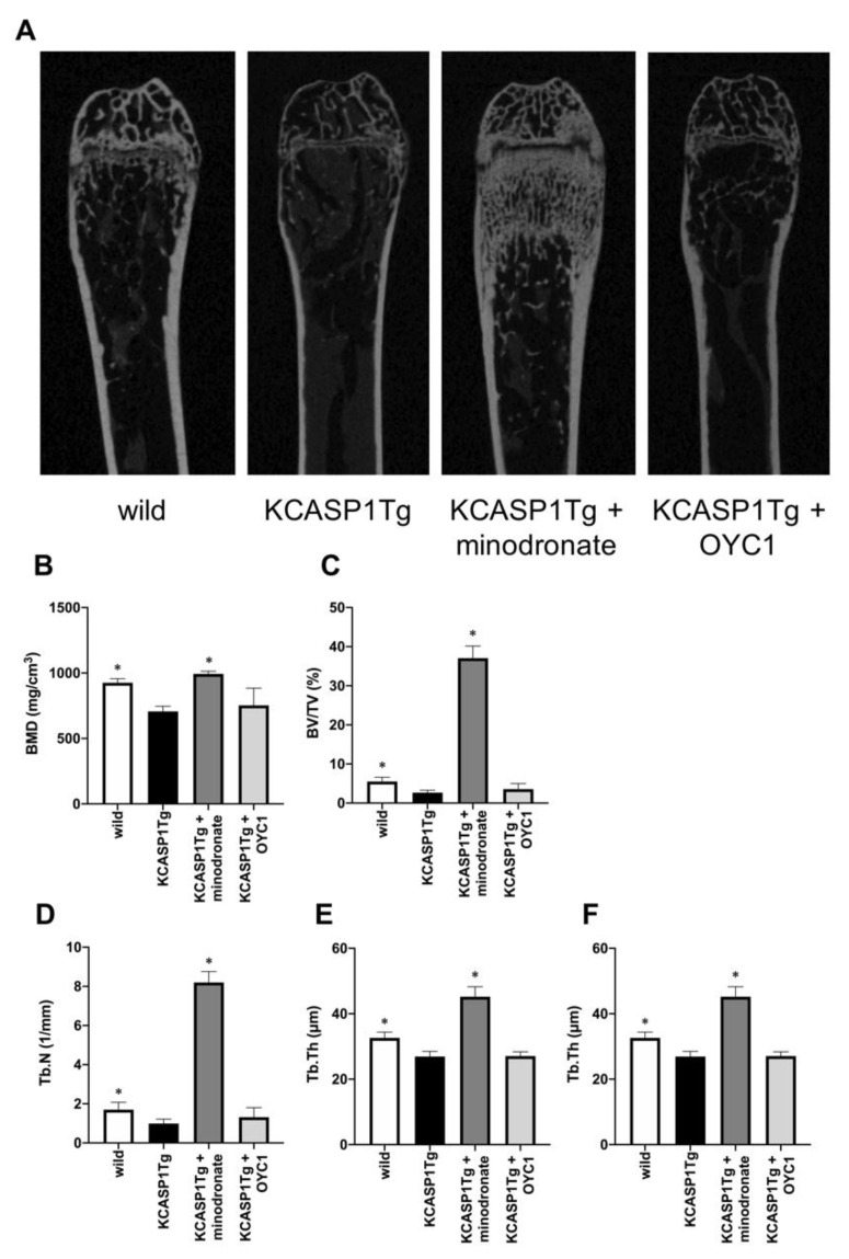
Figure 3.
Analysis of the trabecular bone structure by µCT. The trabecular bone structure was evaluated by µCT in KCASP1Tg mice, treated with or without minodronate or OYC1 (n = 4 per each group) (A). BMD was decreased in KCASP1Tg mice and a marked augmentation of bone mass was observed in minodronate-treated KCASP1Tg mice (B). The bone volume per tissue volume (BV/TV) was reduced by 52% in KCASP1Tg mice compared to wild-type littermates and was significantly restored by minodronate (C). The trabecular number (Tb.N) was decreased in KCASP1Tg mice and was substantially recovered after treatment with minodronate, while OYC1 showed a moderate ameliorative effect (D). The distance between the trabeculae, the travecular separation (Tb.Sp), was increased (E), and the trabecular thickness (Tb.Th) was significantly decreased in KCASP1Tg mice (F). Minodronate induced a significant recovery. All data were expressed as mean ± SD. *; p < 0.05, compared to KCASP1Tg mice by Mann–Whitney test.

