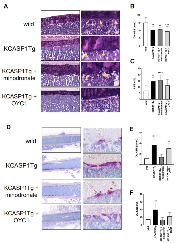
Figure 5.
Histomorphometric evaluation by HE and TRAP staining. HE staining (left panel × 100 and right panel × 400) of the femoral diaphysis in wild-type, KCASP1Tg, and treated KCASP1Tg mice (A). The number of osteoblasts per bone surface (Ob.N/BS) was significantly decreased (B) and the eroded surface per bone surface (ES/BS) was increased in KCASP1Tg mice compared to wild-type littermates. ES/BS was not improved in both minodronate- and OYC1-treated KCASP1Tg mice (C). Ob.N/BS and ES/BS were quantified in three random parts of the HE section in each sample (×100, n = 3). TRAP staining (left panel scale bar is 300 µm, right panel × 400) of the femoral diaphysis in wild-type, KCASP1Tg, and treated KCASP1Tg mice (D). Osteoclast number per bone surface (Oc.N/BS, (E)) and osteoclast surface per bone surface (Oc.S/BS, (F)) were analyzed in three random parts of the TRAP section in each sample (×100, n = 3). Oc.N/BS and Oc.S/BS were significantly increased in KCASP1Tg mice compared to wild-type mice. Minodronate treatment reduced both Oc.N/BS and Oc.S/BS, whereas OYC1 ameliorated only Oc.S/BS (E,F). All data are expressed as mean ± SD. *; p < 0.05, **; p < 0.01, ***; p < 0.001, ****; p < 0.0001 compared to KCASP1Tg mice by Mann–Whitney test.

