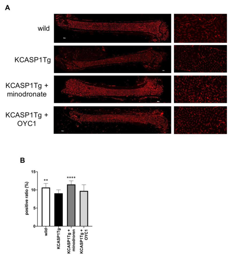
Figure 6.
Blood flow evaluation in the femur by MECA-32 staining. In order to evaluate the blood flow in the femur, we performed fluorescent immunostaining with MECA-32, a vascular endothelial cell marker. In KCASP1Tg mice, the diameter of the blood vessels in the bone marrow was decreased (left panel scale bar is 300 µm, right panel × 100, (A)). We calculated the blood vessel area per bone marrow area (positive ratio) in three randomly selected locations (×100, n = 3 per group). The ratio was decreased in KCASP1Tg mice compared to wild-type littermates, while minodronate significantly improved the vascular area (B). **; p < 0.01, ****; p < 0.0001 compared to KCASP1Tg mice by Mann–Whitney test.

