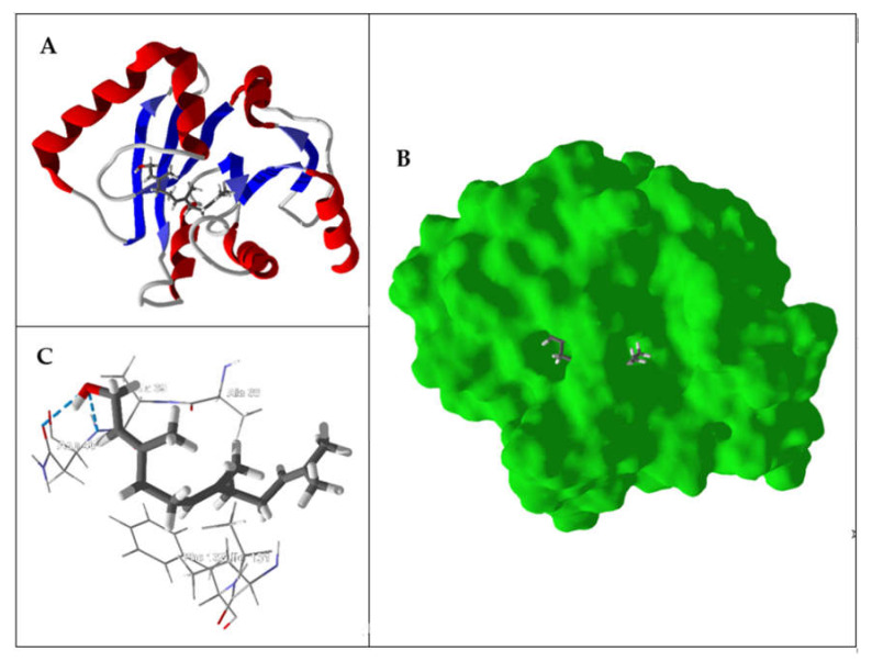
Figure 4.
Lowest-energy docked pose of (E,E)-farnesol with SARS-CoV-2 ADP ribose phosphatase (PDB: 6W02). (A) Ribbon structure of the enzyme and the docked ligand. (B) Solid structure of the enzyme showing (E,E)-farnesol in the binding cleft. (C) Amino acid residues in proximity to the docked (E,E)-farnesol (hydrogen bonds are indicated with blue dashed lines).

