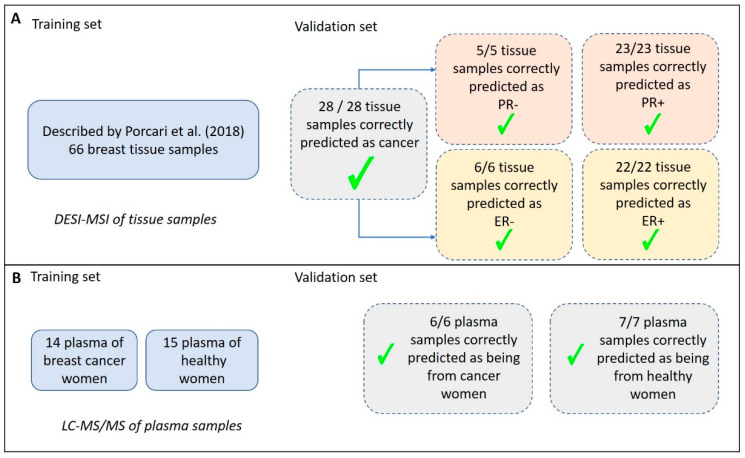
Figure 2.
Summary of the classification predictions of breast carcinoma tissue and plasma samples. (A) Results obtained for tissue analysis using DESI-MSI (Desorption-Electrospray-Ionization—Mass Spectrometry Imaging). A previously validated model for classification of samples was described by Porcari et al. [28], with 66 breast cancer samples compared to normal breast tissue, and it was used here as a test set. In the validation set, all the NST (no special type) and special type tissue samples were correctly classified as being cancer and as having +/- Progesterone Receptor (PR) and +/- Estrogen Receptor (ER). (B) Describes the results obtained for plasma analysis using LC-MS/MS (Liquid Chromatography—tandem Mass Spectrometry). The test set was composed of 29 plasma samples and resulted in average accuracies of 99.8% (positive ion mode) and 99.2% (negative ion mode) based on 100 cross-validations. In the validation set, including 30% of the samples, all the plasma samples were correctly classified as being cancer or not.

