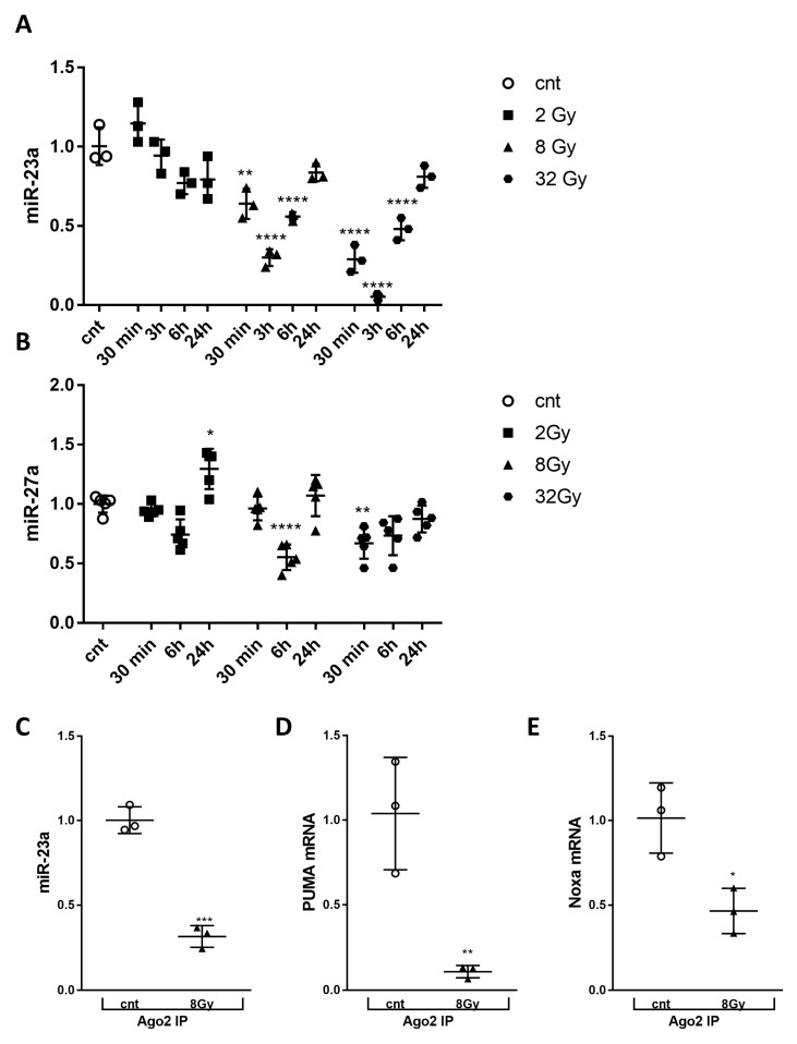
Figure 4.
Irradiation decreased levels of cellular miR-23a-3p, and miR-23a-3p, PUMA, and Noxa mRNAs within the RNA-induced silencing complex. Neurons collected at 30 min, 6 h, and 24 h after 2, 8, and 32 Gy irradiation. Total RNA was used for qPCR analysis. qPCR quantification of miR-23a-3p (A) and miR-27a-3p (B) levels in a representative experiment. The experiment was repeated three times. N = 3/group in each experiment for miR-23a-3p, n = 5/group in each experiment for miR-27a-3p, with 2 technical replicates. Data represent the mean ± SD of one-way ANOVA and Tukey post-hoc analysis * p < 0.05, ** p < 0.01, *** p < 0.001, **** p < 0.0001 vs. control RCN. Neurons were collected 3 h after 8 Gy treatment, subjected to RIP with Ago2 antibodies, and samples used for qPCR analysis. qPCR quantification of miR-23a-3p (C), Puma (D) and Noxa (E) levels in precipitates after RIP in a representative experiment. The experiment was repeated two times, n = 3/group in each experiment with 2 technical replicates. Data represent the mean ± SD. Statistical significance assigned by one-tailed t-test, *** p < 0.001 versus control, n = 3/group.

