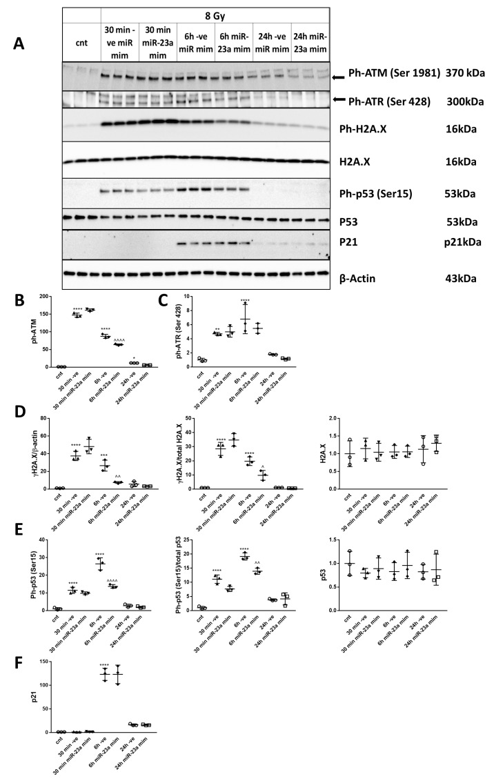
Figure 6.
miR-23a-3p attenuates DNA damage response and p53 activation in primary cortical neurons following irradiation. RCNs were transfected with miR-23a-3p mimics, and negative control mimics before irradiation. Neurons were collected at 30 min, 6 h, and 24 h after 8 Gy irradiation. Whole-cell lysates were separated by SDS-polyacrylamide gel and immunoblotted with antibodies against Ph-ATM (Ser1981), Ph-ATR (Ser428) (approximately 300 kDa), γ-H2A.X (Ser139), H2A.X, Ph-p53 (Ser15), p53, p21 and β-actin (representative image). Image of a representative experiment (A). Protein levels were quantified by densitometry, normalized to β-actin and the parent proteins for γ-H2A.X and Ph-p53, and presented as fold change compared with untreated control levels (B–F). The experiment was repeated 3 times with similar results, n = 3/group in each experiment. Data represent mean ± SD. Statistical significance assigned by one-way ANOVA and Tukey post-hoc analysis, * p < 0.05, ** p < 0.01, *** p < 0.001, **** p < 0.0001 vs. control RCN, ^^ p < 0.01, ^^^^ p < 0.0001 vs. negative control mimic transfected cells (-ve mim) RCN.

