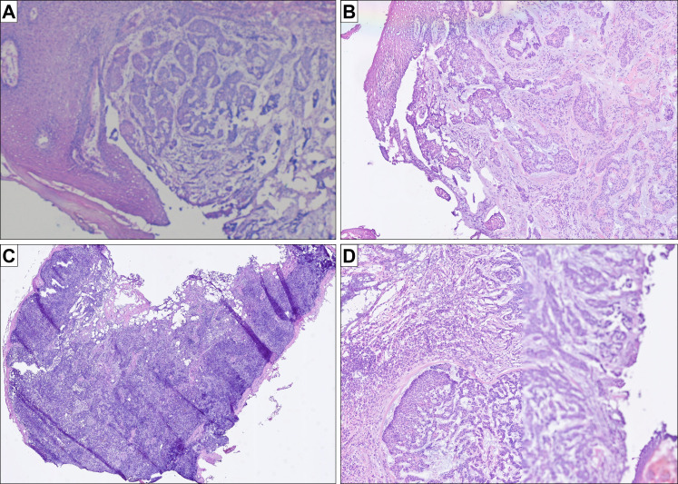
Figure 2.
Histology images and potential pitfalls and advantages of WSI (all figures represent original images without postprocessing). (A) routine quality of H&E frozen sections slides using robotic microscopy and the iPath system (basal cell carcinoma); (B) routine quality of the same H&E frozen section slide using uPath; (C) case with many folds, in which the IMS managed to correctly focus on the thin evaluable areas without the need of further manual readjustment (sentinel lymph node); (D) case with a focus issue in one of the serial sections: while the left half of the tissue is focused correctly, the right half is out of focus; the second serial section was entirely focused (basal cell carcinoma of B). IMS, image management software; WSI, whole slide imaging.

