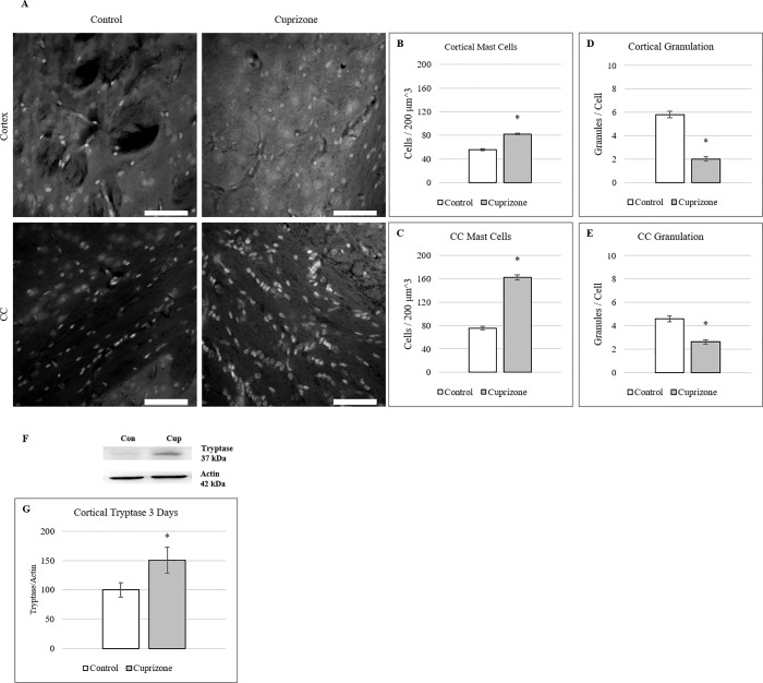
Fig 5. Mast cell activation occurs at 3 days of cuprizone administration.
(A) Toluidine Blue stained mast cells in cortical and corpus callosum tissue after 3 days of cuprizone administration. (B-C) Quantification of mast cell presence shows significant increases in both cortical and corpus callosum tissue after 3 days of cuprizone administration. (mean ± s.e.m.). n = 6 (D-E) Measures of mast cell degranulation shows a decrease in stained mast cell granules in both cortical and corpus callosum tissue after 3 days of cuprizone administration. n = 30 (mean ± s.e.m.). Scale bar = 50 μm, Asterisk denotes significance (p = < 0.05) F) Western blot analysis of cortical Tryptase at 3 days of cuprizone administration. Representative blots of Tryptase and Actin in mice after 3 days of cuprizone administration. G) Densitometry of western blot shows significant increase in Tryptase levels compared to controls. Data are expressed as a percentage of control with the highest control set at 100%. n = 3. Asterisk denotes significance (p = < 0.05).

