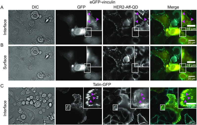
Fig 2. Co-labeling of focal adhesion markers vinculin or talin with HER2.
Direct interference contrast (DIC) and wide field fluorescence images of SKBR3 cells seeded on glass-bottom dishes. The cells were either transfected with eGFP-vinculin (A and B) or transduced with talin- GFP (C) to label focal adhesion spots (GFP signal, magenta arrow heads). HER2 was labeled by biotinylated anti-HER2 affibody coupled to strept-QD (HER2-Aff-QD). (A) The focus was adjusted to the basal side of the cells at the interface with the substrate. Images were acquired using a 63x objective. (B) Similar images as in (A), but with the focus adjusted to apical side of the cell. (C) Images of SKBR3 cells with labeled HER2 and transfected with talin-GFP. Images were acquired using a 40x objective. Colors in merged images: yellow for GFP and cyan for HER2-Aff-QD. Scale bars: 20 μm and 10 μm for the insets.

