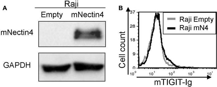
Figure 5.
Murine Nectin4 does not bind murine TIGIT. (A) Overexpression of murine Nectin4 (indicated as mNectin4) on Raji cells. Western blots were performed with antimurine Nectin4 AB and expression was compared with Raji cells expressing empty vector (indicated as empty). Staining for GAPDH was used as a loading control. (B) FACS staining of Raji cells transfected either with an empty vector as control (gray histograms), or with murine Nectin4 (black histograms). Cells were stained with murine TIGIT-Ig. Figures show one representative experiment out of three performed. Graph depicting the mean fluorescence intensity values of the stainings appears in online supplementary figure 4 FACS, fluorescence-activated cell sorting.

