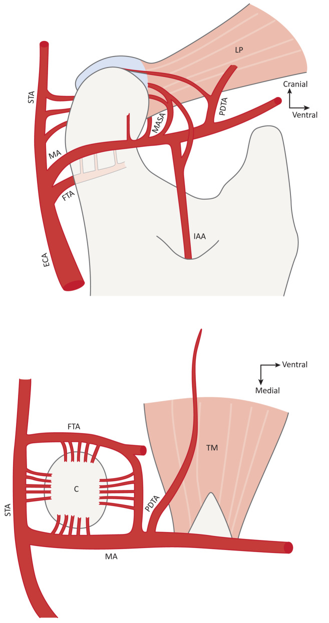
Figure 6. Schematic representation of vascular supply to mandibular condyle according to Toure (61). The figures are slightly modified. Top: Medial view of condylar region illustrating the arterial circle surrounding the articular process. Bottom: Cranial aspect of arterial supply to the condyle (STA: superficial temporal artery; LP: lateral pterygoid muscle; MA: maxillary artery; MASA: masseteric artery; PDTA: posterior deep temporal artery; FTA: facial transverse artery; ECA: external carotid artery; IAA: inferior alveolar artery; TM: temporal muscle; C: condyle). For further details on condylar vascular anatomy, see the publication of Toure (61).

