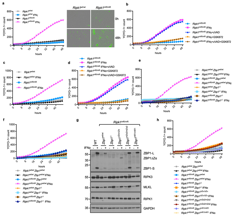
Extended Data Figure 5. IFNα and IFNγ induce death of Ripk1mR/mR cells by inducing Zα-dependent ZBP1 activation and RHIM1-mediated downstream signalling.
a, Cell death measured by YOYO-1 uptake in Ripk1wt/wt or Ripk1mR/mR MEFs treated with IFNγ (1,000 u ml−1) for 48 h and IncuCyte images of Ripk1wt/wt or Ripk1mR/mR MEFs before and 48 h after IFNγ (1,000 u ml−1) treatment. YOYO-1 staining is shown in green. b-f, h, Graphs depicting cell death assessment by YOYO-1 uptake in MEFs with the indicated genotypes treated with combinations of IFNγ (1,000 u ml−1), IFNα (50 ng ml−1), Z-VAD-FMK (20 μM) and GSK’872 (3 μM) for 48 h. g Immunoblot analysis of total lysates from MEFs with the indicated genotypes stimulated with IFNγ (1,000 u ml−1) for 24 h. Representative data in panel a (n = 7), b (n = 5), c (n = 3), d (n = 3), e (n = 7), f (n = 3), g (n = 2) and h (n = 3). Panels (a-f, h) show mean values from technical triplicates (n = 3). GAPDH was used as a loading control for immunoblot analysis. For gel source data, see Supplementary Fig. 1.

