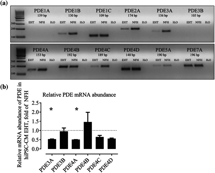
FIGURE 1.

(a) Qualitative RT‐PCR of different PDE‐isoforms (35 cycles). NFH: non‐failing heart. (b) Quantitative RT‐PCR. Relative mRNA abundance of PDE3 and 4 isoforms in hiPSC‐CM EHTs, normalized to NFH, mean ± SEM, n = 5 for NFH and hiPSC‐CM EHTs (ΔΔCT). P < .05, significantly different from NFH samples; t test for unpaired samples. Note, higher sensitivity allowed detection of PDE3B in quantitative PCR (Figure 1b) but not in qualitative PCR (Figure 1a). This figure is related to Figure S1
