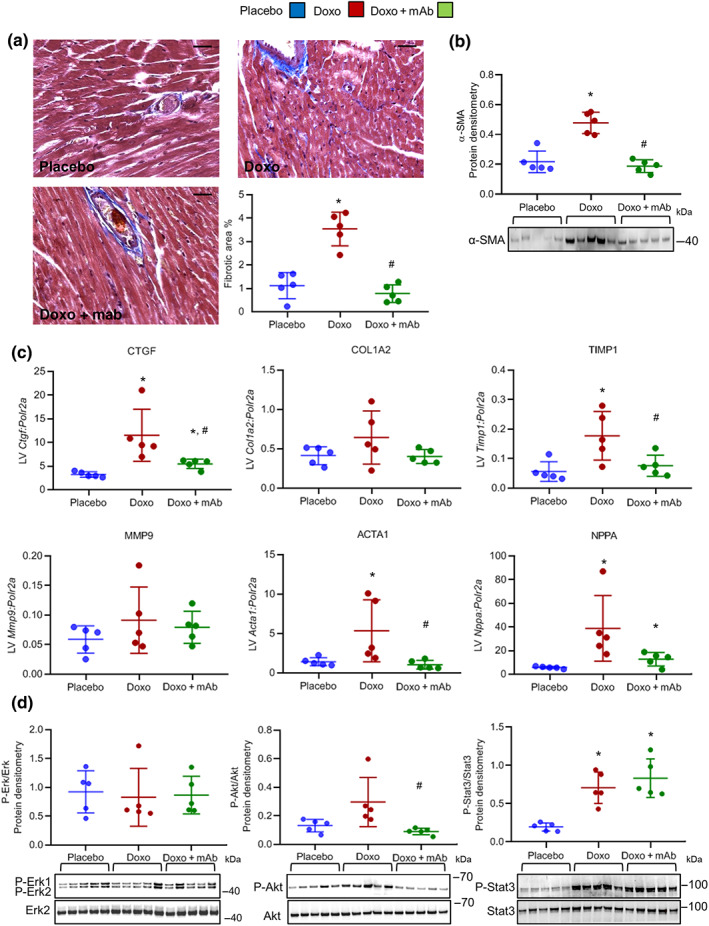
FIGURE 3.

The agonist mAb exerts cardioprotective effects towards doxorubicin‐induced cardiotoxicity. (a) Representative images and quantification analysis of Masson's trichrome staining on hearts of placebo, Doxo, and Doxo + mAb mice at Day 35 after onset of doxorubicin treatment. Bars: 25 μm. (b) Western blot quantitative protein densitometry (above) and representative image (below) of α‐smooth muscle actin (α‐SMA) protein in heart tissues (n = 5 per group). (c) Gene expression analysis of connective tissue growth factor (Ctgf), collagen type I α 2 chain (Col1a2), TIMP metallopeptidase inhibitor 1 (Timp1), metallopeptidase 9 (Mmp9), α‐actin (Acta1), and atrial natriuretic peptide (Nppa) in the three cohorts of animals (n = 5 per group). Polr2a was used as reference gene for the expression data normalization. (a–c) For mice treatments, see Figure S1. (d) Western blot analysis was performed on heart lysates from mice of three treatment groups (placebo, Doxo, and Doxo + mAb; n = 5 per group). Doxorubicin (15 mg·kg−1) was injected i.p. as a single dose, and mAb (5 mg·kg−1) was administered 24 hr before doxorubicin. Mice were killed 48 hr after doxorubicin treatment. Quantitative protein densitometry (above) and representative images (below) of phosphorylated ERK1/2, Akt, and STAT3. ERK2 was used as loading control in all western blots. *P < .05, significantly different from placebo; # P < .05, significantly different from Doxo group
