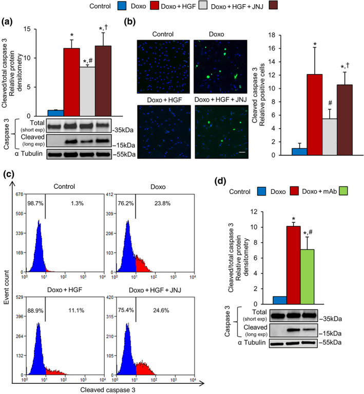
FIGURE 5.

MET activation attenuates doxorubicin‐induced apoptosis in H9c2 cardiomyoblasts. (a–c) H9c2 cells were untreated (control) or treated with doxorubicin (Doxo, 25 μM), Doxo + HGF (0.5 nM), and Doxo + HGF + JNJ (500 nM). Cleaved caspase 3 apoptotic marker was quantified by (a) western blots, (b) immunofluorescence (cleaved caspase 3 green and DAPI blue; bar: 100 μm), and (c) flow cytometry. (d) H9c2 cells were pretreated with 100‐nM mAb instead of the natural ligand, HGF. The level of cleaved caspase 3 protein was determined by western blot (densitometry on the top and representative image on the bottom). Total and cleaved caspase 3 proteins (a, d) were evaluated using two different exposure times; α tubulin was used as loading control in all western blots. All experimental data were obtained from five independent experiments. *P < .05, significantly different from control; # P < .05, significantly different from Doxo; † P < .05, significantly different from Doxo + HGF
