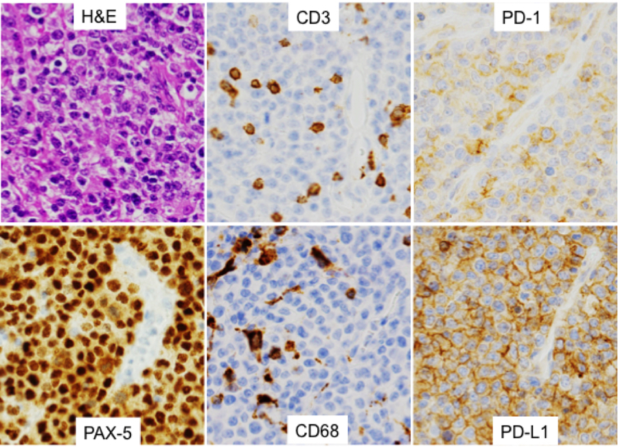
Figure 1.

Representative photomicrographs. A) Hematoxylin-and-eosin (H&E)-stained section of a left frontal lobe mass from a 28-year-old male with HIV. The lymphoma cells are large with round to oval nuclei, coarse vesicular chromatin, distinct nucleoli, and scant cytoplasm. B) PAX5 immunostain highlights the lymphoma cells, confirming their B-cell origin. C) CD3 immunostain highlights tumor-associated small T lymphocytes. In this case, T cells represent approximately 15% of the overall specimen cellularity. D) CD68 highlights tumor-associated macrophages, in this case also representing approximately 30% of the cellularity. In this case, the lymphoma cells express both PD-L1 (E) and PD-1 (F). All images were obtained at 400x magnification.
