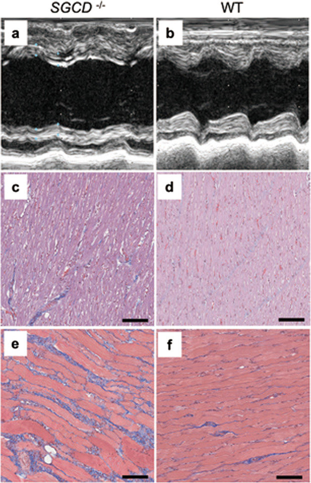
Fig. 8. Cardiac and skeletal muscle pathology recapitulated in SGCD−/− progeny.

The SGCD−/− progeny at 9 weeks (a, c, d) showed the pathological features of the cardiac and skeletal muscle observed in the cloned founder SGCD−/− animals. These features included dilated LV cavities during the systolic phase detectable by echocardiography (a, b), interstitial edema as indicated by gaps between the cardiac muscle cells (c, d), and fibrotic regeneration in the skeletal muscle (e, f). b, d, f WT pig. HE (c, d) and Masson’s trichrome (e, f) staining. Scale bars, 100 µm.
