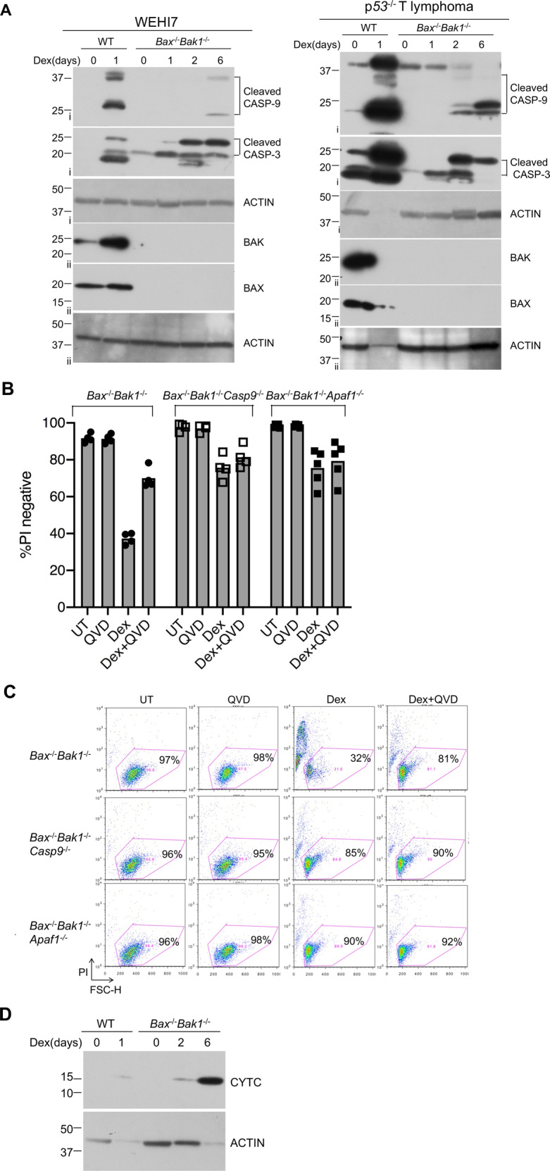
Fig. 2. Dexamethasone can induce caspase activation in the absence of BAX and BAK1.

a WEHI7 cells (left panel) and p53−/− lymphoma cells (right panel) from each genotype (Bax+/+Bak1+/+ and Bax−/−Bak1−/−) were treated with 1 µM Dex for indicated times. Cell lysates were analyzed by western blot with antibodies to cleaved Caspase-3, cleaved Caspase-9, BAK, BAX, and ACTIN. Data show results of one of two independent experiments. Roman numerals to the left of blots (i–ii) indicate the membrane probed. b Mutating Caspase-9 or Apaf1 genes prevented Dex-induced PI uptake in Bax−/−Bak1−/− WEHI7 clonal lines. In the lines mutant for Caspase-9 or Apaf1, addition of QVD did not increase the percentage of PI-negative cells. Independent WEHI7 cell clones from each genotype (Bax−/−Bak1−/−, Bax−/−Bak1−/−Casp9−/− and Bax−/−Bak1−/−Apaf1−/−) were treated with 1 µM Dex and/or 10 µM QVD for up to 6 days. Cells were harvested, resuspended in PBS containing PI, and analyzed by flow cytometry. Data show one of two independent experiments using four or five independent clonal lines. c Dot plots of independent clones from experiments shown in b; numbers represent the percent of PI-negative cells in a total of 10,000 cells analyzed per condition. d Dexamethasone induces cytochrome c release in a Bax/Bak1 independent manner in WEHI7 cells. Cytoplasmic extracts from WT and Bax−/−Bak1−/− WEH7 cells, which were treated with 1 µM DEX for 0 to 6 days, were subjected to western blot analysis, with antibody specific for cytochrome c (CYTC) and ACTIN. Results are from one of three independent experiments.
