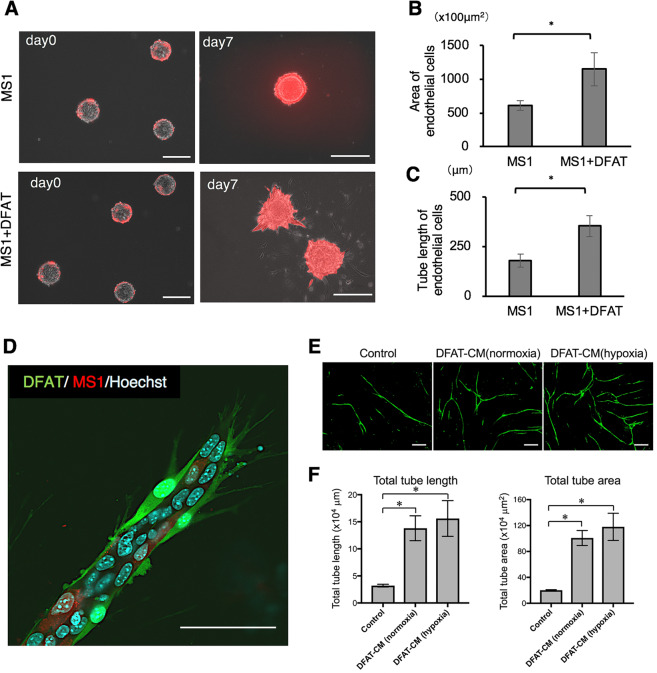
Figure 4.
DFAT cells promote endothelial cell tube formation in vitro. (A) Representative images of collagen beads with mCherry-labelled MS1 cells in the collagen gel 3D culture with DFAT cells on days 0 and 7. Scale = 200 μm. (B,C) Quantification of the area (B) and the tube length (C) of DsRed-positive MS1 cells. (D) Fluorescence microscopic image of the tubules of DsRed-labelled MS1 cells with GFP-labelled DFAT cells. Scale = 200 μm. (E,F) The effect of DFAT cell conditioned media on tube formation ability in human umbilical vein endothelial cells were examined. Representative photomicrographs of tube-like structures in each group. Scale = 200 μm (E). Quantification of total tube length and total tube area in each group (F). Values are represented as mean ± SD. *p < 0.05.

