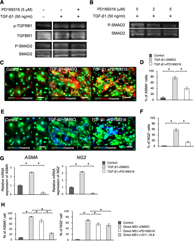
Figure 6.
Smad2/3 activation plays an important role in differentiation of DFAT cells into mural cells. (A,B) Western blotting analysis for phospho-TGFBR1 and phospho-Smad2 expression in DFAT cells. Protein expression of phospho-TGFBR1, phospho-Smad2 in DFAT cells in the presence of TGF-β1 and a Smad2/3 inhibitor PD169316 (A). Effects of PD169316 in different doses on phospho-Smad2 in DFAT cells (B). (C–G) The changes of pericyte marker expressions in DFAT cells on adding DMSO or PD169316 into the culture medium was examined with TGF-β1. Immunofluorescence staining of GFP labelled DFAT cells with ASMA (C) and NG2 (E) in each of the groups. Scale = 30 μm. The percentage of ASMA-positive (D) and NG2-positive (F) cells. The gene expression of ASMA and NG2 in DFAT cells in the presence of TGF-β1 and PD169316 (G). (H) The changes of pericyte marker expressions in DFAT cells in the presence of a Smad2/3 inhibitor or a TGF-β1-neutralizing antibody, in co-culture with MS1 cells. The percentage of ASMA or NG2-positive cells (H). Values are represented as mean ± SD. *p < 0.05.

