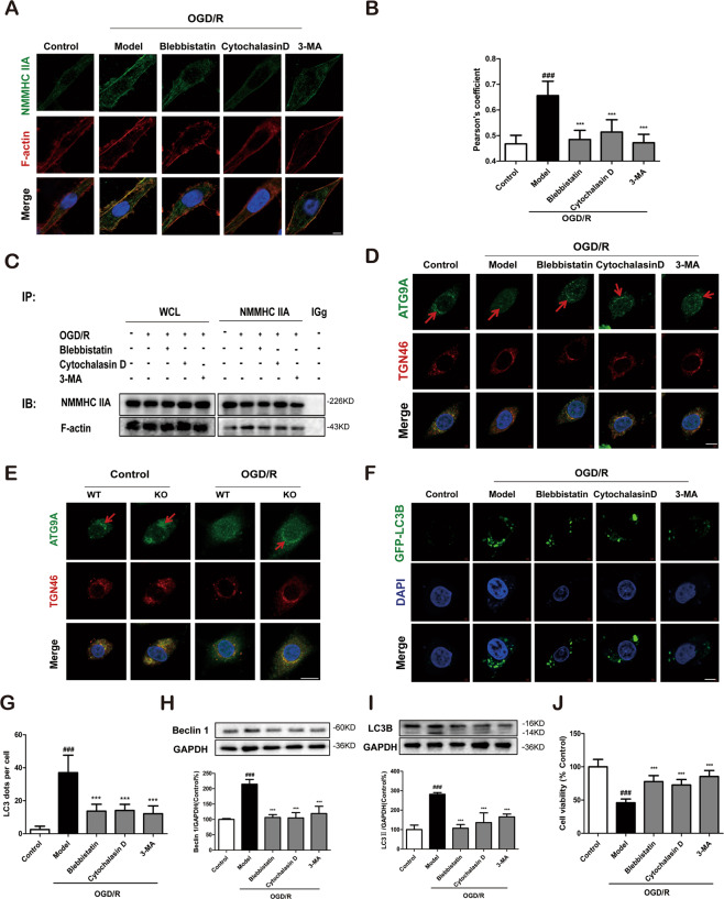
Fig. 5. Inhibition of the NMMHC IIA–actin interaction alleviates ATG9A trafficking and neuronal autophagic cell death during OGD/R in PC12 cells.
PC12 cells were treated with 6 h of OGD and then reoxygenated for 6 h. a Confocal microscopy was used to detect NMMHC IIA (green), F-actin (red), and DAPI (blue). Bar: 2 µm. b Colocalization of NMMHC IIA with F-actin was evaluated by Pearson’s coefficients. c Co-IP was used to detect protein interactions between actin and NMMHC IIA. d ATG9A (green), TGN46 (red), and DAPI (blue) were detected by confocal microscopy. Bar: 2 µm. e ATG9A (green), TGN46 (red), and DAPI (blue) were detected by confocal microscopy in NMMHC IIA knockout PC12 cells. Bar: 2 µm. f, g PC12 cells were transfected with GFP-LC3B plasmid, and EGFP-LC3B puncta was detected after OGD/R treatment. Bar: 2 µm. h, i Beclin 1 and LC3B expression are depicted by immunoblotting. j Cell viability was evaluated by MTT assay upon OGD/R treatment. Results are expressed as the mean ± SD, n = 3. ##P < 0.01 vs. control group; **P < 0.01, *P < 0.05 vs. Model group.

