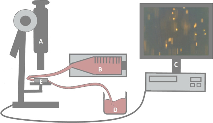
Figure 1.
Experimental setup of the flow-adhesion assay. Capillaries (E) were monitored microscopically (A). Flow rates of RASF-containing suspensions were regulated by a syringe pump (B). The pump was connected to the capillaries by a tube. Another tube was connected to a collection vessel (D) after passing through the capillaries. Synovial fibroblast migration was evaluated by three video sequences per setting (C).

