
Fig. 3.
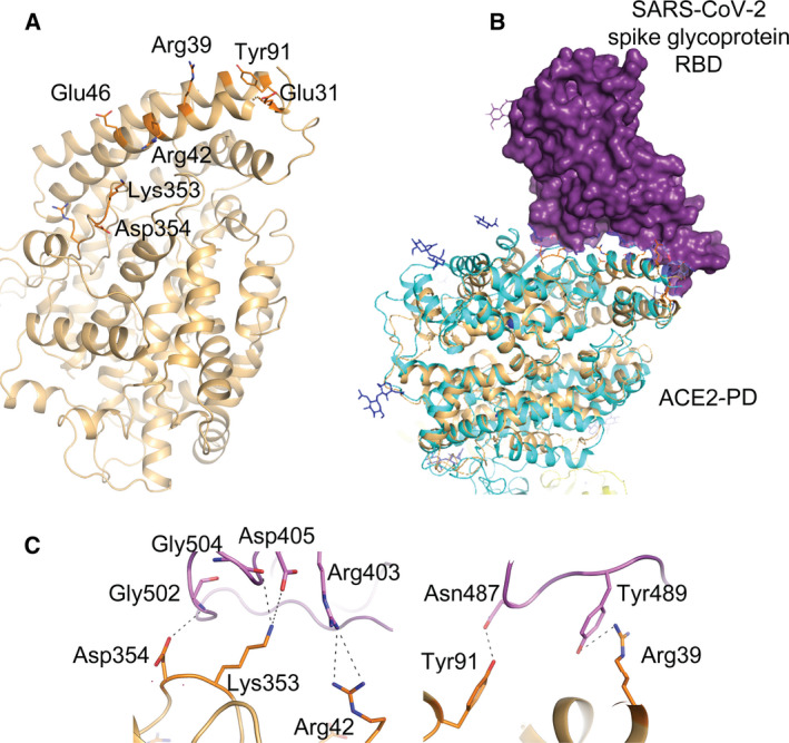
Members of proteobacteria express a peptidyl peptidase with similarity to the PD of human ACE2. (A) The predicted structure of peptidyl peptidase from Proteobacteria bacterium. The structure was predicted using Phyre2 server. The amino acid residues of the putative‐binding site of the S‐protein are numbered. (B) Alignment of the predicted structure of the bacterial ACE2‐PD‐like peptidyl peptidase (ochre) with the 3D structure of ACE2‐PD (cyan) in complex with the RBD of the SARS‐CoV‐2 S‐protein (purple). (C) Possible interactions between amino acid residues of bacterial ACE2‐PD‐like peptidyl peptidase (orange) and those of RBD of the SARS‐CoV‐2 (Pink).