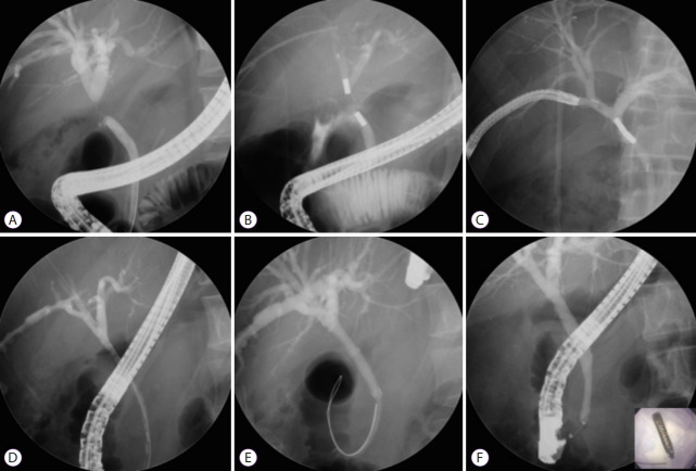
Fig. 4.
Magnetic compression anastomosis for a post-cholecystectomy stricture. (A) Common hepatic duct stricture that occurred after laparoscopic cholecystectomy. (B) One magnet was moved through the percutaneous transhepatic biliary drainage (PTBD) tract, and a second magnet was delivered via the common bile duct using a duodenoscope. Magnet approximation was successful but the distance between two magnets was long. However, the two magnets moved closer to each other because of magnetic power, and magnet approximation was successful. (C) The approximated magnets were removed using percutaneous transhepatic cholangioscopy via the PTBD tract and endoscopic retrograde cholangiopancreatography scope. (D) Cholangiogram showing the recanalized tract after magnet removal. (E) A retrievable, fully covered self-expandable metal stent (FCSEMS) was inserted for 6 months (exchanging every 3 months). (F) Finally, formation of a new fistula was confirmed after the removal of the indwelling FCSEMS. The bottom right color photograph shows the removed FCSEMS.

