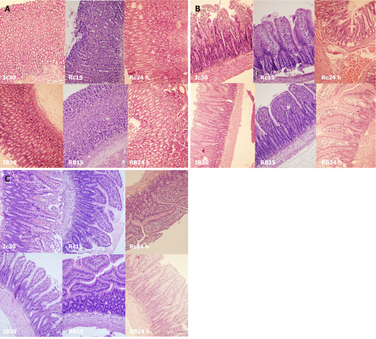
Figure 12.
BPC 157 effect on gastrointestinal lesions microscopy presentation [stomach, duodenum, ileum]. A: Stomach (HE staining, × 20), Controls: Enhanced capillary congestion and erythrocytes extravasation present at the end of 30 min portal triad obstruction (PTO) ischemia period (Ic30). With reperfusion, as seen at 15 min of reperfusion the lesion progressed to enhanced capillary congestion, mild lifting of surface epithelial layer from lamina propria (Rc15). At the 24 h of reperfusion appears enhanced capillary congestion in lamina propria with erythrocytes showing ischemic changes (Rc24 h). BPC 157: Mildly villous surface, rare erythrocytes with ischemic changes present at the end of 30 min PTO ischemia period (IB30). With reperfusion, glandular architecture preserved at 15 min of reperfusion (RB15). At the 24 h of reperfusion appears only mild capillary congestion in lamina propria is seen. Architecture of glands is preserved (RB24 h). B: Duodenum (HE staining, × 20), Controls: Loss of villous architecture, loss of surface epithelium, edema of lamina propria present at the end of 30 min PTO ischemia period (Ic30). With reperfusion, the lesion progressed as seen with edema of lamina propria with extravasation of erythrocytes, some of them with ischemic changes, and focal loss of surface epithelium at 15 min of reperfusion (Rc15). At the 24 h of reperfusion appear enhanced capillary congestion, focal erythrocytes showing ischemic changes (Rc24 h). BPC 157: Mild capillary congestion in lamina propria is seen with edema of lamina propria. Extravasation of erythrocytes, elevation of surface epithelium from lamina propria at the end of 30 min PTO ischemia period (IB30). Villous architecture is preserved. Edema of lamina propria with extravasation of erythrocytes and reparatory changes of epithelium. Mild capillary congestion in lamina propria is seen at 15 min of reperfusion (RB15). At the 24 h of reperfusion villous architecture is preserved. Edema of lamina propria (RB24 h); C: Ileum (HE staining, × 20), Controls: Loss of villous architecture, enhanced elevation of surface epithelium from lamina propria with loss of surface epithelium, edema of lamina propria, marked capillary congestion present at the end of 30 min PTO ischemia period (Ic30). With reperfusion, edema of lamina propria with extravasation of erythrocytes. Some of them with ischemic changes. Mild lymphocytic infiltrate. Focal elevation of surface epithelium from lamina propria, at 15 min of reperfusion (Rc15). At the 24 h of reperfusion appear enhanced capillary congestion, focal erythrocytes showing ischemic changes (Rc24 h). BPC 157: Mild capillary congestion in lamina propria is seen with mild edema of lamina propria. Mild elevation of surface epithelium from lamina propria. Some lymphocytes infiltrating lamina propria at the end of 30 min PTO ischemia period (IB30). Villous architecture is preserved, edema of lamina propria with extravasation of erythrocytes and mild lymphocytic infiltrate is seen at 15 min of reperfusion (RB15). At the 24 h of reperfusion mild capillary congestion in lamina propria is seen, villous architecture is preserved, edema of lamina propria (RB24 h).

