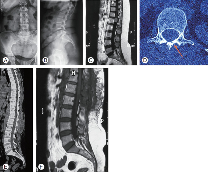
Fig. 5.
(A, B) Preoperative radiographs showing normal spinal alignment and (C) IDEM tumor at L1 level on MRI (later proved to be a meningioma on histopathology). (D) Healing of osteotomy site is seen in axial CT film on one side at 6-month follow-up (arrow). (E) Maintained sagittal alignment on sagittal CT film. (F) No tumor recurrence observed on MRI. IDEM, intradural extramedullary; MRI, magnetic resonance imaging; CT, computed tomography.

