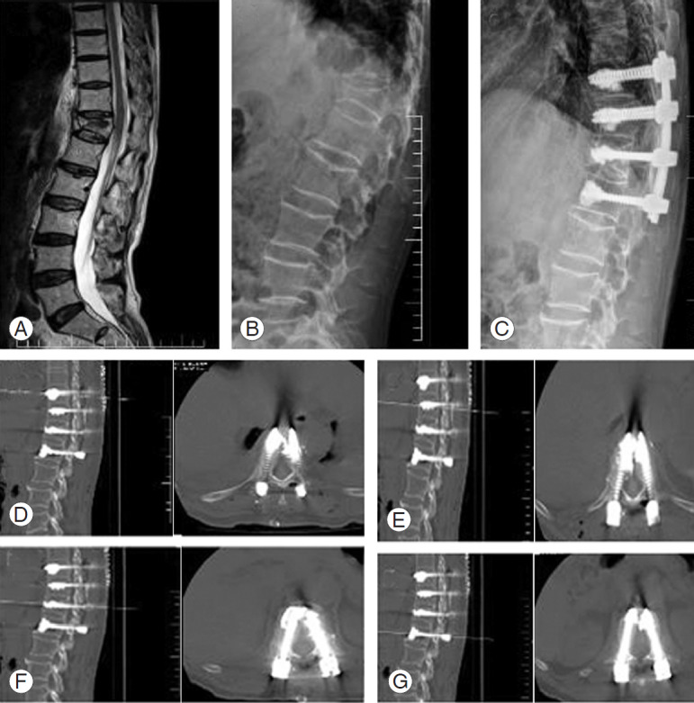
Fig. 3.

(A) Sagittal T2 magnetic resonance imaging of a 75-year-old male who presented to us with severe back pain not responding to conservative management and showing an osteoporotic T12 fracture with marrow edema. His dual absorption X-ray absorptiometry scan suggested osteoporosis with t-value of −3.5. (B, C) Preoperative and postoperative X-rays showing fracture fixation using the modified technique. (D–G) Computed tomography scan images showing bicortical fixation with cement in the vertebral body. Bicortical fixation could not be performed at the fractured vertebra.
