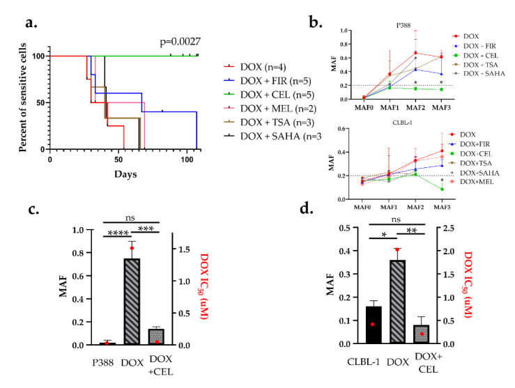
Figure 4.
Celecoxib prevented the emergence of Pgp-mediated drug resistance in both P388 and CLBL-1 cells. (a) Kaplan–Meier curves of cell cultures treated with the indicated combinations. Cells were considered resistant at MAF ≥ 0.2. DOX (red) in combination with firocoxib (blue), celecoxib (green), meloxicam (pink), trichostatin-A (brown), and SAHA (black) were treated in 9 sequential treatment cycles. (b) Quantitative evaluation of multidrug resistance during the course of various treatments in P388 and CLBL-1 cells. MAF was measured after every third treatment cycle (MAF1–3). (c,d) Relation of MAF and drug sensitivity. MAF (patterned bars) and DOX IC50 values (red squares) of parental (sensitive), DOX-treated, and DOX + CEL-treated (c) P388 cells, and (d) CLBL-1 cells, measured after 9 sequential treatment cycles (at around day 100). Statistical analysis was performed on MAF values, * p < 0.05, ** p < 0.01, *** p < 0.001, **** p < 0.0001, ns: not significant.

