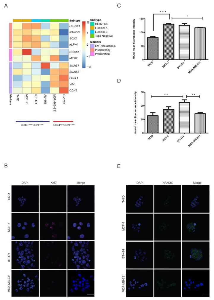
Figure 3.
Luminal CD44−/low/CD24−/low cells show a similar expression profile of pluripotency markers. Comparison of mRNA expression of markers for pluripotency, EMT/metastasis and proliferation for CD44high/CD24−/low and CD44−/low/CD24−/low cells, performed using qRT-PCR. (A) Heat map of genes with opposite expression patterns between CD44high/CD24−/low and CD44−/low/CD24−/low for pluripotency and EMT. Each row represents an RNA transcript; each column represents a cell line. CD44−/low/CD24−/low cells from luminal cell lines overexpressed pluripotency markers (Supplementary Figure S3). Representative image and analysis of immunofluorescence for MKI67 (B,C) and NANOG (D,E) expression in mammospheres from CD44−/low/CD24−/low and CD44high/CD24−/low cells. Flow cytometry sorted cell populations were maintained as spheres for 14 days and stained with DAPI (blue), NANOG (green) and MKI67 (magenta). Spheres were observed under fluorescence microscope. Data represent the mean ± SD (n = 3) of three independent experiments; * p < 0.05, ** p < 0.01 and *** p < 0.001. Scale bar = 100 μm.

