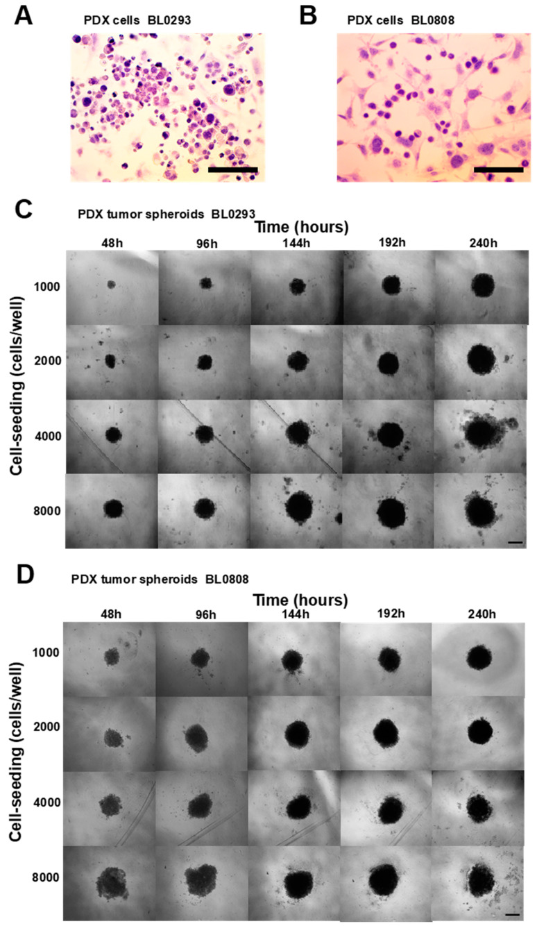
Figure 1.
Bright-field microscopic images of patient-derived xenograft (PDX) tumor cells BL0293 (A) e BL0808 (B) stained with hematoxylin and eosin (tumor cells purplish/bluish-colored and fibroblasts pinkish-colored); 40× magnification, scale bar = 100 μm. Phase-contrast microscopic images of PDX tumor spheroids BL0293 (C) and BL0808 (D) at different cell-seeding concentrations, cultured for 10 days; 10× magnification, scale bar = 500 μm.

