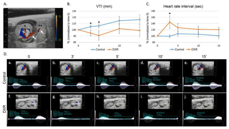
Figure 1.
Blood flow in the umbilical cord. (A) On day E12.5 of pregnancy, an umbilical cord artery of one embryo was viewed by Color Doppler mode; presentation of an embryo (*) with its adjacent placenta (**) and the umbilical cord (arrows) in which the acquisition was taken. Following a short stabilization period, a baseline arterial blood flow was recorded and quantified. Then, mice were injected with either doxorubicin (DXR; 8 mg/kg, n = 7, n = number of imaged arteries, one embryo per pregnant mouse) or saline (control; n = 6). The arterial blood flow was monitored continuously by pulse-wave (PW) Doppler mode for 15 min, recorded and analyzed at various time points post injection by analyzing the (B) velocity time integral (VTI); and (C) heart rate interval. (D) Representative PW Doppler photos of the analyzed time points. Analysis of each PW Doppler cine loop was performed. Values of post-treatment imaging were normalized according to pretreatment imaging values of each mouse (defined as 100%). Saline-injected mice were standardized to 100% as a reference to DXR-injected mice. Data are expressed as mean ± standard error of mean (SEM). Individual comparisons were made using a Student’s t-test. (*) P value of 0.05 was considered statistically significant.

