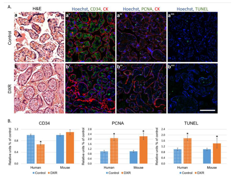
Figure 4.
Histology of placentae. (A) Human placentae from women diagnosed with breast cancer during pregnancy (week 27–35) and treated with EPI (n = 7) were compared with human placentae from chemo-naïve women (control n = 10). Representative pictures of control- (a–a‴) or EPI-exposed (b–b‴) human placenta, stained for the following: H&E (a,b); CD34 (neovascularization a′,b′); PCNA (proliferating cell nuclear antigen; a″,b″); pan-cytokeratin (positive control for staining; CK a′,a″,b′,b″); or TUNEL (apoptosis a‴,b‴). Florescence images were photographed using a LSM-510 confocal laser-scanning microscope. Offset calibration of the photomultiplier was performed with sections stained with secondary antibodies only. Bar = 100 µm. (B) Florescence pattern was imaged in human and in dissections of placentae from pregnant female mice treated with either DXR (n = 52, from 4 pregnant mice) or saline (n = 21, from 4 pregnant mice). The average number of PCNA positive cells, TUNEL positive cells, or CD34 blood vessels were automatically analyzed by FIJI software, and compared with control samples (defined as 1). Data are expressed as mean ± SEM. Individual comparisons were made using a Student’s t-test. (*) P value of 0.05 was considered statistically significant.

