Figure 1.
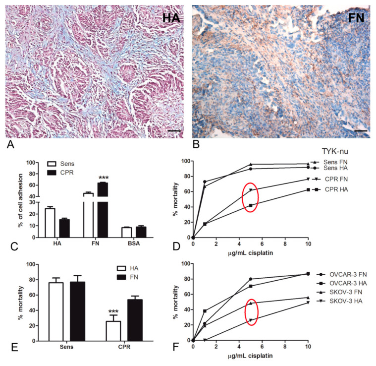
(A,B) Hyaluronic acid (HA) and fibronectin (FN) presence at the tissue level in human high-grade serous ovarian cancer (HGSOC). (A) Histochemical staining with Alcian Blue highlighted the HA distribution in HGSOC tissue sections; in particular, the staining was visible in tumor-associated stroma. Magnification, 200×; scale bar, 50 μm. (B) Immunohistochemical assay for the detection of FN at the tissue level showed a strong presence of FN in the tumor stroma and around blood vessels. A streptavidin–biotin–peroxidase system with 3,3′-Diaminobenzidine tetrahydrochloride (DAB) was used. Magnification, 200×; scale bar, 50 μm. (C–E) HA matrix maintained the chemoresistance of the ovarian cancer cell lines. Cisplatinum-sensitive (Sens) TYK-nu and cisplatinum-resistant (CPR) TYK-nu were stained with a fluorescent dye (Fast DiI) and seeded onto a HA- or FN-coated 96-well plate. The percentage of adhesion was interpolated using a calibration curve generated through the lysis of an increasing number of labeled cells. On HA, the adhesion of Sens TYK-nu was favored compared to that of CPR, whereas on FN, CPR appeared to be more adhesive than sensitive ones; both cell types preferentially adhered to FN. The fluorescence intensity was measured using Infinite200 (Tecan Group Ltd., Männedorf, Switzerland) (C). By evaluating the percentage of mortality after 72 h of treatment with 5 µg/mL of cisplatinum, we observed that hyaluronic acid was increasingly able to maintain chemoresistance as compared to FN. Cell viability was evaluated with an ELISA reader (O.D., 450 nm) (D,E). A killing assay was also performed with the OVCAR-3 and SKOV-3 cell lines, observing a similar behavior to that of the TYK-nu cells (F). *** p < 0.001.

